
Engineering Cellular Microenvironments
The local microenvironment of cells is known to exhibit a wealth of regulating cues in affecting cell behaviour. In order to better investigate these processes, to maintain the naturally occurring state of cultivated cells in lab, and to elaborate new states for analytical, biotechnological and therapeutical applications, there is a need for advanced cell culture systems. These engineered biomaterial scaffolds are intended to allow a tight control of key parameters of the cellular microenvironment.
In a matrix engineering approach we design and build functional microenvironments for specific biomedical and biotechnological situations, including stem cell niches, cancer progression, wound healing as well as microbial consortia and biofilms.
Matrix Reconstitution
Biomolecular engineering of cell scaffolds of mammalian tissues can be performed through the in vitro reconstitution of supramolecular structures of extracellular matrix components such as proteins, proteoglycans, and glycosaminoglycans. Among the biomacromolecules utilized for that purpose, collagen is distinguished by its abundance in the mammalian organism, its importance for the connective properties of tissues, and a wide variety of specific interactions with nearly 50 different molecules including glycosaminoglycans (e.g. heparin), proteins (e.g. fibronectin), and numerous growth factors. We study the dynamics and structural polymorphism of in vitro reconstituted collagen matrices. Similar approaches are used for fibrin matrices as well. Based on a thorough analysis, variations in composition, morphology, mechanics as well as orientational alignment are utilized to design scaffolds in 2D and 3D arrangements.
Similar approaches and technologies can be used to engineer 3D (bio-)polymer-based matrices to study and manipulate microbial cell growth and intercellular communication. In there other materials classes (e.g. agarose, poly(vinyl alcohol) and other parameter ranges (pore size, stiffness) are implement to control microbial cell behavior.
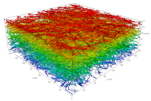
Reconstituted 3D collagen network imaged by laser scanning microscopy. Image: F. Ullm
Selected publications:
- Biomaterials 52:367 (2015) Link
- J Materials Chemistry B 3:8902 (2015) Link
- Adv Healthcare Mat 5:1861 (2016) Link
Cytokine Presentation
Cytokines and morphogens have been identified as essential cell fate triggers. We engineer in vitro microenvironments that allow to control cytokine presentation in both a temporal and spatial fashion. Topics like local gradients as well as the mode of presentation (i.e. bound vs. non-bound) are studied in order to build biopolymer matrices with biomimetic characteristics to guide cell behaviour in different physiological and pathological situations.
Selected publications:
in vitro Models of Wound Healing, Cancer Invasion and Stem Cell Niches
Based on our biomimetic models of the extracellular matrix using well-defined, advanced 3D scaffolds based on fibrillar collagen and biohybrid microstructured polymers we set-up various local microenvironments of early and late stages of dermal wound healing, breast and melanoma cancer cell invasion into the surrounding tissue, and the hematopoietic stem cell niche. We explore important regulating features of these microenvironment in controlling cell behaviour. In this context, we modulate parameters such as the composition, topology, stiffness and interface architecture of the biopolymer matrices, as well as the presentation of cytokines in bound and soluble manner. Cell fate is studied on population and single cell level using state-of-the-art cell analytical techniques including in situ single cell tracking.
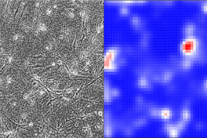
Particle Image Velocimetry analysis of living fibroblasts in 3D collagen networks. Left – phase contrast image, right – respective convergence image. Picture: P. Riedl
Selected publications:
- Sci Reports 6:31951 (2016) Link
- Adv Healthcare Mat 5:1861 (2016) Link
- Adv Biosystems 4:1900220 (2020) Link
Material-Assisted Models of Microbial Consortia and Biofilms
Structured organization of microbial consortia within biofilms as found in nature are envisioned great potential for biotechnological applications, like substrate degradation for chemical precursors, complex biosensing, and production of advanced chemical compounds. As understanding of organizational principles and design criteria of structured microbial consortia is still limited, possible applications still awaiting to be explored and implemented. We investigate how biomaterial engineering can be used to design specific consortia and microenvironmental conditions and mimic naturally occurring states and possible industrial applications. We develop new systems to adjust important microenvironmental parameters and allow for in-depth analysis with high temporal and spatial resolution and a broad range of metabolic relevant parameters.
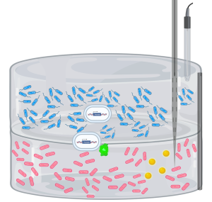
Concept for using biomaterial engineering to construct and analyze spatially and compositionally defined microbial consortia. Picture: C. Danneberg (created using Biorender)
Selected publication:
- Curr Opin Biotechnol 81:102916 (2023) Link
Biosensors
Label free biosensors are desired screening methods in the environmental monitoring, pharmaceutical industry and research settings. The advantage of these techniques lies in their ability to probe biomolecular interactions with high selectivity without the risk of altering the affinity of the analyte molecules. The developments in this field are directed towards more affordable sensors with increased throughput and enhanced selectivity and sensitivity. Other sensor devices rely on functional molecular modification and linking technologies where polymeric materials are highly efficient tools as they allow for a broad variety of chemistries and functionalities by easy tunable synthesis strategies. We develop several new sensor principles and modifications including a new force-based detection technique based on soft colloidal probes, nanoparticles and regenerative sensor surfaces.
Soft Colloidal Probes
Our current focus is set on a new label-free screening technique. As detection principle we use soft colloidal probes (SCP) and their adhesion induced mechanical deformation at ligand / receptor interfaces. We construct various biosensors for easy and straightforward, but still highly sensitive on-site applications. Further direction is the determination of specific cell material interactions via combination of SCP and AFM.
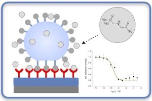
Soft colloidal probe assay for biosensing of glyphosate. Picture: T. Pompe
Selected publications: