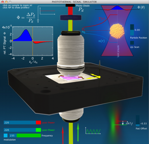
This applet-simulator (M. Selmke) shows the key parts of a typical photothermal confocal microscopy setup: A piezo scanner (black) which can move the sample (purple, blue) which is fixed to it in lateral and axial directions, the heating and the detection laser, the two microscope objectives and the transmission detecting photo-diode. For a detailed account see the publication “Photothermal Single-Particle Microscopy: Detection of a Nanolens“, M. Selmke, M. Braun, F. Cichos, ACS Nano 6, 2741−2749 (2012).
The axial coordinate can be controlled by the particle position slider in the top right corner. Moving the piezo stage has the effect of moving a particle in the sample relative to the fixed focused beams (red and green). A continuous particle position scan can be done by clicking the checkbox.
The laser powers can be adjusted in the bottom left corner. The green heating laser can be modulated with the modulation checkbox above and the modulation frequency may be adjusted. The green heating laser can further be offset relative to the probing beam (controller and illustration in the bottom left).
The resulting PT signal (red/blue) is plotted in the top left corner, while its angular distribution is shown in the very top right corner. Here, also the probe-beam modification is illustrated. The temperature and refractive index profiles around the particle may be shown by clicking on the big particle.

